Revolutionary microscopy technique allows scientists to better understand amyloid-based diseases such as Alzheimer’s and Parkinson’s
02/03/2019 / By Edsel Cook
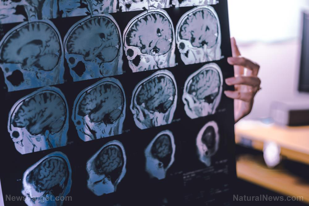
A new breed of powerful microscopes has enabled British researchers to make huge strides in the study of amyloid diseases. According to a Science Daily article, cryo-electron microscopes have enabled new discoveries about the proteins that cause Alzheimer’s, Huntington’s, and Parkinson’s diseases.
These powerful instruments use electrons to view the shape of the samples. They can discern tiny details that are almost as small as an atom.
The University of Leeds (Leeds) have two cryo-electro microscopes. Researchers used them to take high-power snapshots of an amyloid structure.
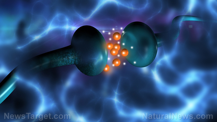
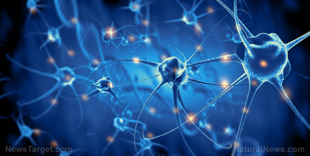
Amyloids are abnormal proteins that accumulate in important organs like the brain. Excess amounts of amyloids form harmful structures that interfere with the healthy function of the organ. Eventually, they lead to serious diseases.
Photos of amyloid structures are few and far between. Images that are of good quality are even rarer. The high-quality 3D images taken by the Leeds researchers are considered to be a huge boon in the study of amyloids.
The images could provide vital clues on the method by which amyloids created their aggregates. They could also show how the harmful proteins increase the risks of an amyloid disease. (Related: The truth about amyloid plaque and its connection to Alzheimer’s disease.)
Cryo-electron microscopes produce crystal clear photos of amyloid structures
The Leeds researchers published their findings in the journal Nature Communications. Their paper showed that the amyloid aggregates formed twisted fibers of considerable length.
The amyloid fibers are made out of beta-2 microglobulin. This protein is normally found in a healthy immune system. However, it could also assemble amyloid structures that can end up inside important body parts or organs.
If these amyloid aggregates end up in the badly-damaged kidneys of patients undergoing long-term dialysis, they can cause severe pain. When they find their way to the joints, they will trigger osteoarthritis.
The photos and 3D structures taken by the Leeds team will be of great interest to organizations that are looking for ways to treat amyloid diseases. For the past 60 years, these study groups have been improving their techniques and understanding of the proteins.
In the past, they used early electron microscopes that produced blurred photos of amyloids. Now, the latest cryo-electron microscopes can provide crystal clear images of amyloid structures.
“We’ve used cryo-electron microscopy not only to uncover the shape and structure of amyloid proteins, but also how they grow and intertwine with each other like the stands in a rope to form larger assemblies,” explained Leeds lead researcher Sheena Radford.
A better understanding of Alzheimer’s disease and other amyloid diseases
Radford explained that the cryo-electron microscope images showed every kink and point on the proteins that made up the amyloid structure. Researchers could develop therapeutic compounds that can either lock down those points or disrupt the connections that made up the fiber.
Furthermore, her team also discovered how the amyloid proteins grow and tie themselves together. They likened the process to how rope makers would entwine numerous strands until an entire rope was formed.
Earlier, less clear images of these amyloid structures portrayed them as simple ladder shapes. But the vastly improved quality of cryo-electron microscope images revealed that the aggregates were much more complex structures than previously believed.
Furthermore, the new findings could be used to compare and contrast the protein structures of various amyloid diseases in individual patients. The Leeds researchers believe that the amyloid structures involved in Alzheimer’s disease is somewhat different from the one associated with Parkinson’s disease. Diagnosis of these related but different diseases could be made much more accurate.
Learn about natural ways to protect yourself against Alzheimer’s and other amyloid diseases at Alzheimers.news.
Sources include:
Tagged Under: Alzheimer's disease, Amyloid, amyloid proteins, breakthrough, microscope, microscopy, Parkinson's Disease, research, technology breakthrough